A 3D CT scan of your teeth, jaws and nerves, so implants and complex treatment are planned on real measurements, not estimates.
A quick, painless 3D scan that shows bone, roots and nerves in detail a flat X-ray cannot, taken only when it changes your plan.

 1000+ Google reviews

 Excellent on Trustpilot
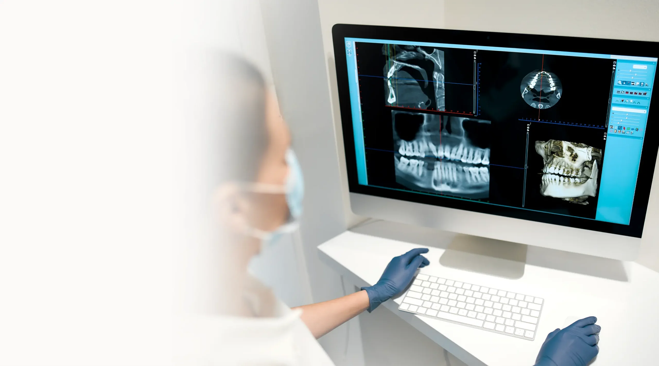
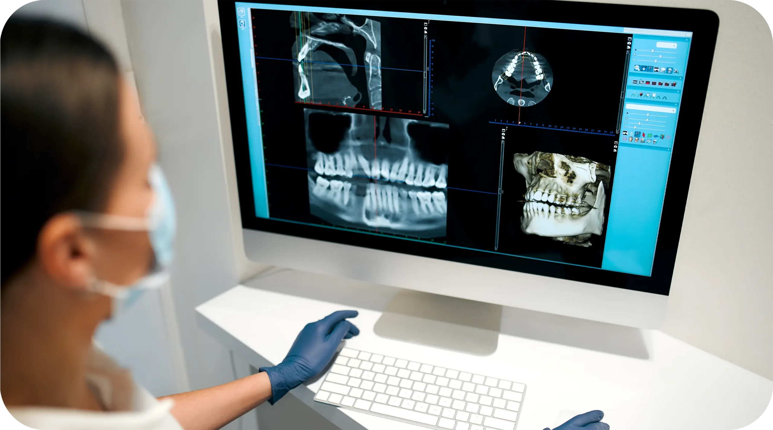
A 3D CT scan shows your jawbone, teeth and nerves in three dimensions, so treatment is planned on measurements, not estimates.
Also called a CBCT scan (cone beam computed tomography), it builds a three-dimensional image from a single rotation around your head. Your dentist can turn it, slice through it and measure it, which a flat X-ray simply cannot do.
A standard X-ray compresses your jaw into one flat image. Structures overlap, distances are distorted, and the width of the bone does not show at all. For a filling, that rarely matters. For an implant, a difficult extraction or a root canal on a complex tooth, it matters a great deal.
The scan shows the height, width and density of the bone, the exact path of the nerve in the lower jaw, the position of the sinuses in the upper jaw, and the shape of every root. Problems that stay hidden on a flat image, such as infection at the tip of a root, often become visible.
Every implant case at HMC is planned on a 3D CT scan. It tells us whether there is enough bone, where the nerve and sinuses sit, and which implant length and diameter fit safely. We place MegaGen implants as our primary system and Straumann as the alternative, and the scan is what lets us choose the right size before surgery rather than during it.
If the bone is too thin or too low, the scan shows that too, and you hear about it at the planning stage, not in the chair.
The scan is taken in the same building where your treatment is planned, and the dentist who reads it is the one who carries out the work, so nothing gets lost between what the scan shows and what is done. If something unexpected appears, such as a cyst, a hidden fracture or bone loss around an old tooth, you hear about it on the day, with the image in front of you.
A 3D CT scan in Albania at HMC is not part of every visit. Many examinations need no imaging at all, and some need only a standard X-ray. When your case does need a scan, your dentist explains why before it is taken, and it appears as a separate line on your written, itemised quotation. You never pay for a scan you did not agree to.
The scanner sends a cone-shaped X-ray beam through the area being examined while an arm rotates around your head once. Software combines the images captured during that rotation into a single 3D model of your teeth, jaws and surrounding structures.
Your dentist tells you what the scan is for and what it should show. You remove glasses, earrings, hairpins, removable dentures and anything else metal around your head and neck, because metal blurs the image. Tell us if you are or might be pregnant. There is no need to fast or prepare in any other way.
You sit or stand in the scanner and rest your chin on a support, and you may be asked to bite gently on a small positioner to keep your jaw still. The arm then rotates around your head. It takes under a minute, it is completely painless, and the machine is open, so it does not feel enclosed. Your only job is to stay as still as possible.
A dental CT scan of this type uses a focused beam and a far lower dose than a medical CT scan of the head. It is higher than a single small X-ray, which is exactly why we only recommend one when the information changes your treatment.
The images are ready almost immediately. Your dentist goes through them with you, showing the bone around the area being treated, the position of nerves and sinuses, and anything unexpected. For implants, measurements are taken directly from the scan to choose the implant and plan its position. You can ask to see any part of the scan again, and your dentist will show you exactly what they are measuring and why.
What the scan shows goes straight into your written treatment plan: whether implants are possible without extra procedures, which teeth can be saved, and what order treatment should follow. If the scan changes the plan, the quotation changes with it, before any treatment starts.
Only when it is needed. A 3D CT scan is not taken by default. Your dentist explains why your case needs one before anything is done, and it appears as its own line on your written quotation. If a recent scan from another clinic is good enough, we use that instead.
A 3D CT scan answers questions a flat X-ray cannot. At HMC it is used mainly for:
Bone height, width and density, plus nerve and sinus positions, measured before surgery to choose the right MegaGen or Straumann implant size and position.
Shows the full shape of curved or extra roots, infection hidden on flat X-rays, and how close an impacted wisdom tooth sits to the nerve.
A recent 3D scan from another clinic is reviewed first. If it covers the right area at good quality, we plan from it instead of repeating it.
A 3D CT scan is usually recommended if you:
If a standard examination is enough to plan your treatment, we will not suggest a scan. You are only charged for imaging your case actually needs.
The scan itself takes under a minute, and it is usually done during your first visit rather than as a separate appointment:
Before the scan, we tell you why it is needed, what it should show and that it is quoted separately. Nothing is taken without your agreement.
You remove glasses, earrings and other metal around the head and neck, then sit or stand still with your chin on a support.
The arm rotates around your head for under a minute. It is painless and open, with no injection or contrast dye.
Your dentist goes through the 3D images with you, showing the bone, roots, nerves and sinuses around the area being treated.
Findings go straight into your written treatment plan and itemised quotation, including implant brand and size where relevant.
The scan is taken in the same building where your treatment is planned and carried out, and the dentist who reads it is the one who treats you. It is only recommended when it changes the plan, and when it does, you see what it shows before you decide anything.
Explore our full range of treatments
No. Both build a 3D image from X-rays, but a dental cone beam scan is designed specifically for teeth and jaws. The beam is focused on a smaller area, the machine is open rather than a tunnel, and the radiation dose is far lower than a medical CT of the head. For dental purposes, a cone beam scan gives the detail needed for implants and complex roots, so there is no reason to use a hospital scanner instead.
The dose from a dental cone beam scan is low, and considerably lower than a medical CT scan. It is higher than a single small dental X-ray, which is why we only recommend one when it changes your treatment. Tell us before the scan if you are or might be pregnant. In that case we would normally postpone it unless there is an urgent clinical reason.
No. You sit or stand still while the arm rotates around your head. Nothing touches you apart from the chin support and, sometimes, a small positioner you bite on gently. The machine is open, so it does not feel enclosed, and the rotation takes under a minute.
The scan itself takes under a minute. With positioning and removing metal items, you are usually in and out within a few minutes. Reading the results takes longer, because your dentist goes through the images with you. That is usually done during the same visit, as part of your examination.
No. It is quoted separately, and that is deliberate. Many examinations need no imaging, so building the cost into every appointment would mean charging people for scans they do not need. When your case does need one, your dentist explains why before it is taken, and it appears as its own line on your written, itemised quotation.
Yes. Every implant case at HMC is planned on a 3D CT scan. It is the only way to measure bone height, width and density accurately and to see exactly where the nerve and sinuses are. Those measurements decide which MegaGen or Straumann implant fits, where it goes, and whether any additional procedure is needed first. Planning implants without a scan means guessing, and we do not do that.
Often, yes. Send it to us before your visit. If it covers the right area, is of good enough quality and nothing significant has changed since it was taken, we plan from it and you do not need a new one. If it is too old, does not show the area being treated, or a tooth has been removed since, we explain why a new scan is needed before taking one.
Remove glasses, earrings, hairpins, necklaces, piercings around the head and neck, and removable dentures or retainers. Metal blurs the image. Bring any previous scans or X-rays you have, and a list of the medications you take. Tell us beforehand if you are or might be pregnant.
Yes, and that is how most patients have it. The examination identifies whether a scan is needed, the scan is taken, and your dentist reviews it with you in the same visit. For international patients, this means the treatment plan and quotation can be ready on the first day of the stay, with treatment starting that week.
Bone width, which a flat X-ray cannot show at all. The exact path of the nerve in the lower jaw. The position of the sinuses. The full shape of curved or extra roots. Infection at the root tip that overlapping structures hide. And the position of impacted wisdom teeth relative to the nerve. In short, it shows depth and the relationships between structures, which is what complex treatment depends on.
If you already have a recent scan, sending it before you travel is the most useful thing you can do. Our clinical team reviews it with your photos free of charge and gives you a preliminary plan. If you have no scan, a local imaging centre can usually take one. Ask us first which area it should cover, so you do not end up needing a second one here.
Send us a few details and our patient coordinators will come back within 24 hours with a treatment plan, timeline, and price. No obligation.